Chapter 6: The consultant’s mother
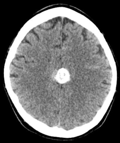
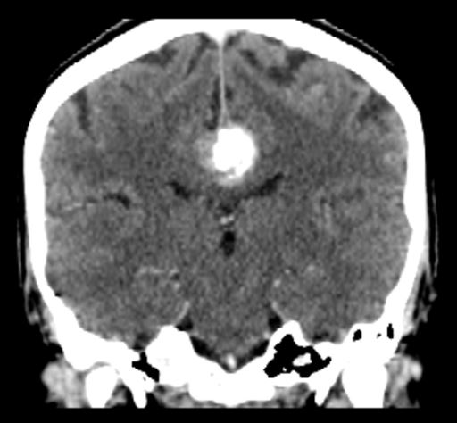
The 72-year-old mother of a consultant radiologist attends the emergency department following a fall at home. She did not lose consciousness, had no neurological deficit or amnesia, her Glasgow coma score (GCS) was 15/15 and she did not vomit. Because she was the mother of a radiologist, she had a CT scan of her head.
-
1. What does the unenhanced CT scan show?
Show Answer
Correct answer:
A calcified parasagittal meningioma.
-
2. What cranial tumours frequently calcify?
Show Answer
Correct answer:
- Oligodendrogliomas (90%)
- Craniopharyngiomas (90%)
- Ependymomas (50%)
- Meningiomas (25%)
- Choroid plexus tumours (25%)
Meningiomas make up about one in five primary central nervous system (CNS) tumours and 90% are benign low grade tumours. On plain skull X-rays they may produce bone erosion, calcification and hyperostosis (excess growth of bone). Meningiomas present with headaches, seizures, motor and sensory symptoms and cranial neuropathies, depending on their site. The most common sites are: (i) parasagittal falx, (ii) olfactory groove, (iii) sella turcica, (iv) sphenoidal ridge, (v) tentorium and (vi) foramen magnum.
![]()
-
3. What neurological signs would you expect from meningiomas at each site?
Show Answer
Correct answer:
- Parasagittal falx: progressive spastic weakness, numbness of legs
- Olfactory groove: anosmia, visual loss and papilloedema (Foster-Kennedy syndrome), frontal lobe syndrome
- Sella turcica: visual field loss
- Sphenoidal ridge: cavernous sinus syndrome (medial sphenoid), exophthalmos and visual loss (middle sphenoid), temporal bone swelling and skull deformity (lateral sphenoid)
- Tentorium: Hydrocephalus, gait ataxia, cranial neuropathies (V, VII, VIII, IX and X at cerebellopontine angle)
- Foramen magnum: Suboccipital pain, ipsilateral arm and leg weakness
The management of meningioma depends on the presence of neurological signs and symptoms. Primary treatment is neurosurgical resection. High grade meningiomas or those that are incompletely resected are treated with post-operative radiotherapy to reduce the rate of relapse.
She was, of course referred to a neurologist and then a neurosurgeon but as neither could find any significant neurological deficit, she was spared surgery to the relief of both her and her son.
Meningiomas are a feature of the hereditary neurofibromatosis (both NF-1 and NF-2). Phakomatoses are a group of familial conditions that cause both cutaneous and neurological manifestations.
-
4. Name as many phakomatoses as you can.
Show Answer
Correct answer:
- Neurofibromatosis (von Recklinghausen’s disease)
- Tuberous sclerosis (Bourneville’s disease)
- von Hippel-Lindau disease (cerebroretinal angiomatosis)
- Sturge-Weber syndrome (encephalotrigeminal angiomatosis)
- Osler-Rendu-Weber syndrome
- Fabry’s disease (angiokeratoma corporis diffusum)
The first three of these are associated with brain tumours.