
Chapter 15: A tired suit
A 34-year-old suit from the city (corporate lawyer) complained of backache and fatigue. On his third visit to the surgery, his GP took a blood test and found that his serum urea was 16 mmol/L and creatinine was 600 mmol/L. He was sent to the emergency department. He had a swollen right testicle, a retroperitoneal mass and bilateral hydronephrosis. He had an ultrasound of his testicle.

-
1. What does the testicular ultrasound show?
Correct answer:
Right testicular ultrasound shows three hypoechoic solid lesions. One has foci of calcification.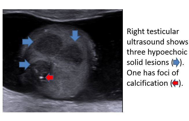
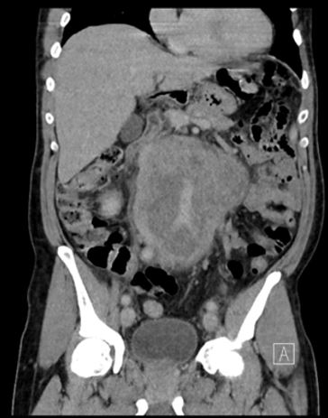
He then had a CT scan.
-
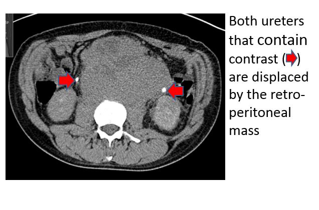
2. What does the CT scan show?
Correct answer:
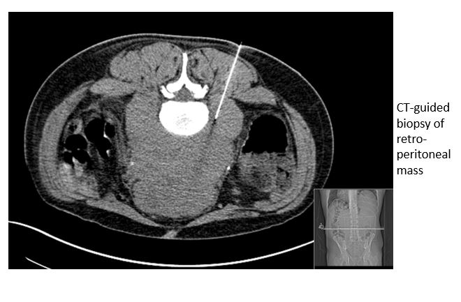
CT shows a confluent retroperitoneal mass encasing the aorta and both ureters that contain contrast are displaced by the retro-peritoneal mass. He has a CT-guided biopsy of retro-peritoneal mass that confirms the diagnosis of seminoma.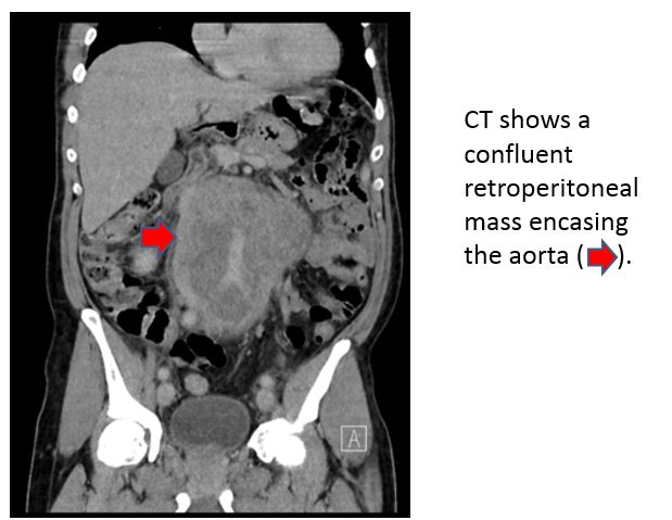
-
3. What serum tumour markers do testicular germ cell tumours produce?
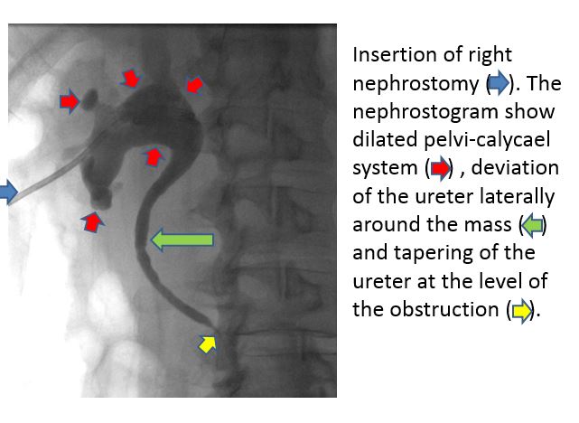
In view of the impaired renal function and hydronephrosis, bilateral nephrostomies were inserted prior to chemotherapy. The right nephrostogram showed a dilated pelvicalyceal system, deviation of the ureter laterally around the mass and tapering of the ureter at the level of the obstruction.
Following the nephrostomies, he was treated with four cycles of BEP chemotherapy for good risk bulky stage IIc seminoma followed by orchiectomy. FDG-PET scan showed no avidity in the residual small volume retroperitoneal mass and he continues on surveillance. Overall testicular cancers represent 1% of all cancers in men and their overall survival is 97%. Since his treatment he has raised over £100,000 for cancer charities.
-
4. What factors increase the risk of developing testicular cancer?